Lifestyle, medical equipments, Technology
What to know about Ultrasound
Oct
What is ultrasound imaging?
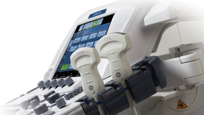
Ultrasound imaging uses sound waves to produce pictures of the inside of the body.
It helps to diagnose the causes of pain, swelling and infection in the body’s internal organs, examine a baby in pregnant women and the brain and hips in infants.
It helps guide biopsies, diagnose heart conditions, and assess damage after a heart attack.
Ultrasound is safe, noninvasive, and does not use ionizing radiation.
This procedure requires little to no special preparation.
Your doctor will instruct you on how to prepare, including whether you should refrain from eating or drinking beforehand.
Leave jewelry at home and wear loose, comfortable clothing. You may be asked to wear a gown.
What are some common uses of the procedure?
Ultrasound examinations help diagnose a variety of conditions and to assess organ damage following an illness.
Ultrasound is used to help physicians evaluate symptoms such as:
-
- pain
-
- swelling
- infection
It is a useful way of examining many of the body’s internal organs, including but not limited to the:
-
- heart and blood vessels, including the abdominal aorta and its major branches
-
- liver
-
- gallbladder
-
- spleen
-
- pancreas
-
- kidneys
-
- bladder
-
- uterus, ovaries, and unborn child (fetus) in pregnant patients
-
- eyes
-
- thyroid and parathyroid glands
-
- scrotum (testicles)
-
- brain in infants
-
- hips in infants
- spine in infants
Ultrasound is also used to:
-
- guide procedures such as needle biopsies, in which needles are used to sample cells from an abnormal area for laboratory testing.
-
- image the breasts and guide biopsy of breast cancer (see the Ultrasound-Guided Breast Biopsy page).
-
- diagnose a variety of heart conditions, including valve problems and congestive heart failure, and to assess damage after a heart attack.
- Ultrasound of the heart is commonly called an “echocardiogram” or “echo” for short.
Doppler ultrasound images can help the physician to see and evaluate the following:
-
- blockages to blood flow (such as clots)
-
- narrowing of vessels
-
- tumors and congenital vascular malformations
-
- less than normal or absent blood flow to various organs
- greater than normal blood flow to different areas which is sometimes seen in infections.
With knowledge about the speed and volume of blood flow gained from a Doppler ultrasound image, the physician can often determine whether a patient is a good candidate for a procedure like angioplasty.
What are the benefits vs. risks?
Benefits
-
- Most ultrasound scanning is noninvasive (no needles or injections).
-
- Occasionally, an ultrasound exam may be temporarily uncomfortable, but it should not be painful.
-
- It is widely available, easy-to-use and less expensive than other imaging methods, imaging is extremely safe and does not use any ionizing radiation.
-
- Ultrasound scanning gives a clear picture of soft tissues that do not show up well on x-ray images, it is the preferred imaging modality for the diagnosis and monitoring of pregnant women and their unborn babies.
- Ultrasound provides real-time imaging, making it a good tool for guiding minimally invasive procedures such as needle biopsies and fluid aspiration.
Risks
- For standard diagnostic ultrasound, there are no known harmful effects on humans
Thank you for your sharing. I am worried that I lack creative ideas. It is your article that makes me full of hope. Thank you. But, I have a question, can you help me?
Your perspective is always so refreshing and insightful.
Hey there You have done a fantastic job I will certainly digg it and personally recommend to my friends Im confident theyll be benefited from this site